Category: Uncategorized
-

Follow My Eye: Using Gaze to Supervise Computer-Aided Diagnosis
One of the most popular trends in medical image analysis is to apply deep learning techniques to computer-aided diagnosis (CAD). However, it is evident now that a large number of manually labeled data is often a must to train a properly functioning deep network. This demand for supervision data and labels is a major bottleneck,…
-
Knee Cartilage Defect Assessment by Graph Representation and Surface Convolution
Knee osteoarthritis (OA) is the most common osteoarthritis and a leading cause of disability. Cartilage defects are regarded as major manifestations of knee OA. However, the cartilage is a thin curved layer, implying that only a small portion of voxels in knee MRI can contribute to the cartilage defect assessment.
-
Recurrent Tissue-Aware Network for Deformable Registration of Infant Brain MR Images
Deformable registration is fundamental to longitudinal and population-based image analyses. However, it is challenging to precisely align longitudinal infant brain MR images of the same subject, as well as cross-sectional infant brain MR images of different subjects, due to fast brain development during infancy.
-
Unsupervised Landmark Detection Based Motion Estimation for Dynamic Medical Images
Dynamic medical imaging in 4D typically requires motion estimation of the organs. With respect to the spatial-temporal information of the 3D volume sampled over multiple time points, one may assess structural and functional property of the target organ. Our early study in CVPR 2020 shows that, with limited number of temporally sampled phase images, one…
-

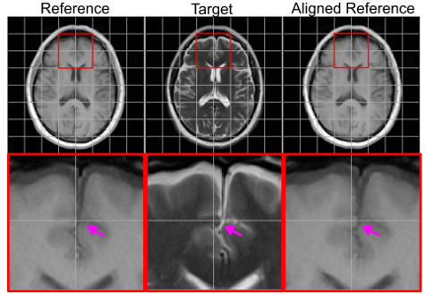
Multi-Modal MRI Reconstruction Assisted with Spatial Alignment Network
In clinical practice, magnetic resonance imaging (MRI) with multiple contrasts is usually acquired in a single study to assess different properties of the same region of interest in human body. The whole acquisition process can be accelerated by having one or more modalities under-sampled in the k-space.